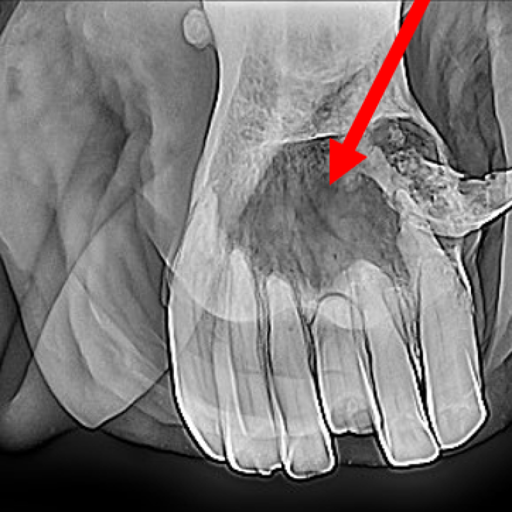
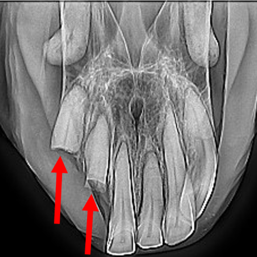
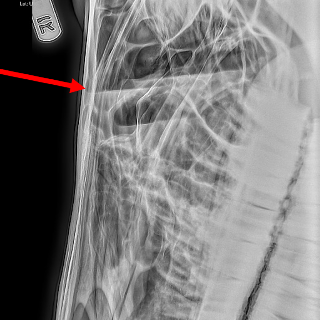
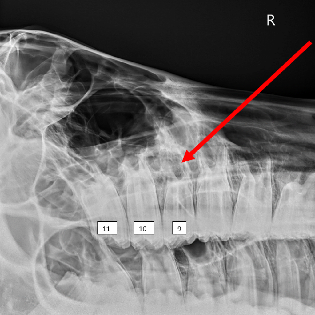
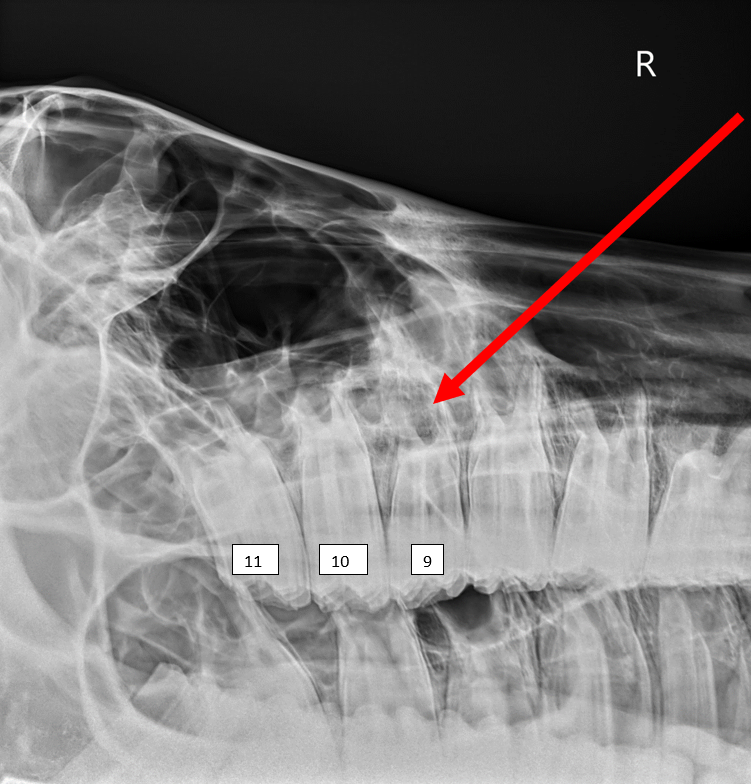
Dental radiographs (Xrays) are a routine procedure for us humans at the dentist’s office, but they are also invaluable for equine dentistry. As equine dentistry has significantly advanced in recent years, improvements in portable radiography equipment have dramatically increased the quality and diagnostic capability of our imaging.
Findings on an oral exam that would indicate the need for dental radiographs include fractured teeth, loose teeth, diseased incisors, or evidence of sinus infection, such as nasal discharge. The portion of the tooth that can be visualized in the mouth is called the clinical crown, but there is significant length of tooth, as well as the roots, hidden beneath the gum line. Radiographs are the best way to evaluate the entire tooth while working in the field. They can identify disease that may not be readily apparent on an oral examination – such as infected or fractured tooth roots. Radiographs can help us decide if a tooth needs to be extracted and plan the best approach for extraction. Serial radiographs are helpful to monitor suspicious teeth over time.
Preparing your horse for dental radiographs is much like preparing your horse for routine dentistry. Appropriate sedation ensures your horse is adequately relaxed and still so that we can capture good images. Depending on the area of interest (sinus, incisor teeth, or cheek teeth), your horse may have his mouth open for images taken at varying angles. We also have an intra-oral plate, similar to what you encounter with your own dentist. Intra-oral plates are great for imaging the incisors, as well as focusing on just few cheek teeth at a time while avoiding overlapping teeth of opposite sides. Our xray equipment allows us to take high-quality images in the field with the images immediately available for review. See below for some interesting Xrays.
