

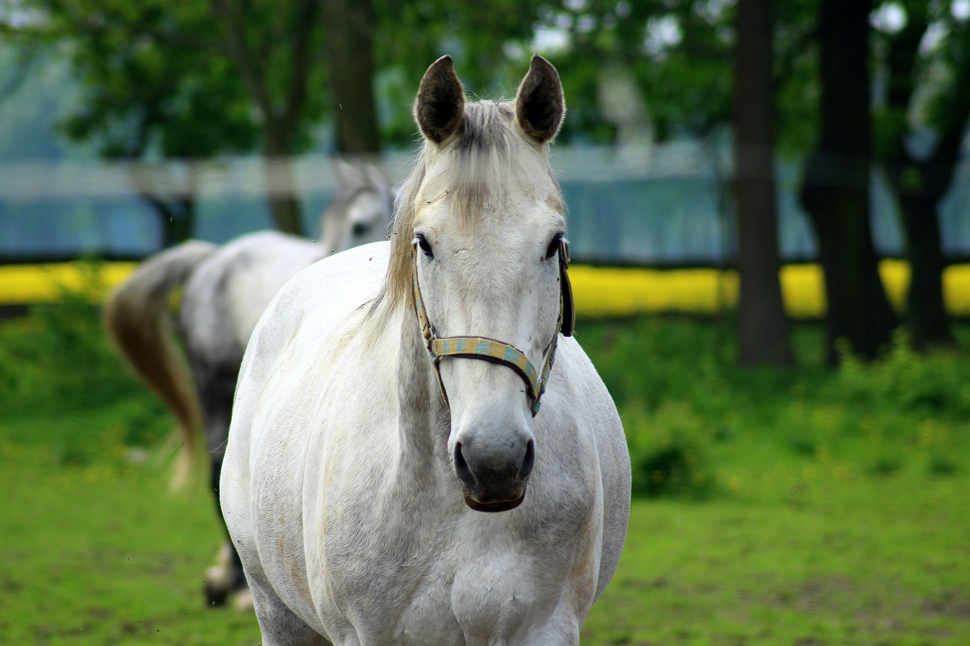
Most horse owners are familiar with equine gastric ulcers, or EGUS (equine gastric ulcer syndrome). Gastric ulcers are extremely common in horses and likely underdiagnosed. Because horses evolved as grazing animals, their stomachs are designed for continual digestion and eating, continually secreting stomach acid. Management of the modern horse often prevents continual grazing, so periods of time without access to forage allow stomach acid to contact the stomach lining, causing ulcerated areas. Common risk factors include periods of time with an empty stomach (such as stalled horses without grazing access), stress (training, showing, travel), and administration of non-steroidal anti-inflammatories (phenylbutazone and banamine).
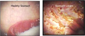
The gold standard for diagnosis is gastroscopy, where your veterinarian will examine the stomach and document findings with photos. Gastroscopy allows us to assign a score of severity to the horse’s ulcers as well as demonstrate healing following treatment. Recent technological advances have made this diagnostic modality available in the field, so gastroscopy can now be performed on the farm instead of trailering in to the clinic.
After you and your veterinarian have decided that gastroscopy is indicated for your horse, the most important part of the gastroscopic exam is patient preparation. Preparation requires fasting your horse for about 20 hours before the exam to ensure your horse’s stomach is empty. If there is food remaining in the stomach, your veterinarian will not be able to perform a complete examination. Frequently we recommend applying a muzzle to your horse during fasting since some horses will ingest shavings or straw. Water must be removed 3 hours prior to gastroscopy.
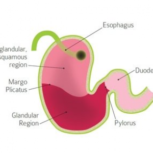
At the appointment, your veterinarian will sedate your horse to allow passage of the scope. The scope is passed up one nostril and to the back of the throat, when your horse will swallow it. During this process, the back of the throat can also be examined. After swallowing, the scope is passed down the esophagus (also examined) and then into the stomach. The upper portion of the stomach is called the squamous/non-glandular portion, based on the cell type composing the lining. The lower portion of the stomach is the glandular portion, where stomach acid is secreted. These 2 areas are divided by a demarcation called the margo plicatus. Ulcers are most commonly found on the squamous portion of the stomach along the margo plicatus. The entire stomach is examined, including all the way to the exit of the stomach (pylorus) and ideally including the beginning portion of the small intestine (duodenum). A score is assigned to your horse based on location of ulcers and the severity of ulceration, ranging from 1-4. Your veterinarian will then work with you to determine the most appropriate treatment for your horse based on this exam. Ideally, the horse is re-scoped following treatment to ensure adequate healing and response to treatment.
MVS is excited to offer gastroscopy to enable appropriate diagnosis and treatment of EGUS, keeping your horse feeling and performing at his best. Contact us today if you’re interested in gastroscopy or would like to learn more.
